| Phone |
| |
| |
| 대표전화 |
1577-7956 |
| 본 사 |
031-457-9187 |
| 대 전 |
042-932-1265 |
| |
|
| Web Site Visitors |
| |
| |
| Today |
1088 |
| Yesterday |
1502 |
| Since 2006 |
2,728,877 |
| |
|
|
|
Lindy O’Clair, Katherine Artymovich, Meagan Roddy and Daniel M. Appledorn
Essen BioScience - Ann Arbor, Michigan
※ IncuCyte Live-Cell Analysis System 을 개발한 Essen BioScience 는 수년전에 Sartorius 사의 일원이 되었습니다. |
Introduction
IncuCyte® Live-Cell Analysis System 을 이용하면 CO2 incubator안에서, 현미경을 이용하여, 살아있는 세포를 직접적으로, 정해진 시간 간격으로,
계속해서 세포변화를 촬영하여, 정량적으로 세포독성 현상을 측정할 수 있는데 IncuCyte CytoTox Reagents 를 이용하여 cytotoxicity 를 측정한다.
세포가 독성 유발물질에 노출되면 여러가지 복잡한 생화학적 변화가 일어나지만 그 중 가장 공통적인 현상이 cell membrane 이 형태적 기능적으로
훼손되는 것, 즉 cell membrane integrity 를 잃게 되는 것이다.
IncuCyte CytoTox Reagents 는 살아있는 cell membrane 을 통과하지 못하지만 세포가 plasma membrane integrity 를 잃게 되면 세포배양액에 있는
IncuCyte CytoTox Reagents 는 세포막과 핵막을 통과하여 핵속의 dsDNA 와 결합하게 된다. IncuCyte CytoTox Reagents 는 DNA 와 결합하지 않은
상태에서는 형광을 발생시키지 않으나 dsDNA 와 결합하게 된 상태에서 빛을 받게되면 종류에 따라 Green, Red 그리고 NIR 과 같은 형광을 발생시키게 된다.
측정하는 세포들에서 발현되는 이 형광을 연속적으로 촬영하고 분석하면 세포의 cytotoxicity 정도를 자동으로 정량적으로 측정이 가능하다.
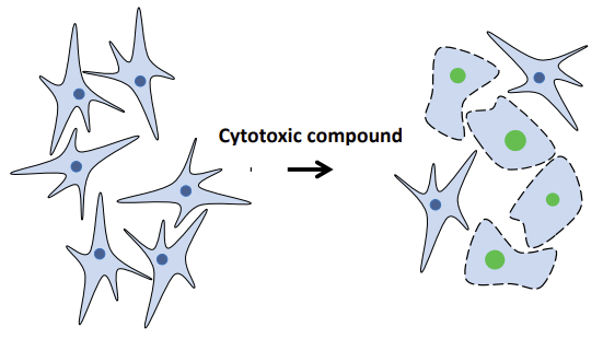
Figure 1. Nuclear staining indicates loss of membrane integrity, a hallmark of cell death. The cell impermeable DNA stain, CytoTox green, stains cell
nuclei only when cells have lost membrane integrity following treatment with a cytotoxic compound. Hallmarks of necrotic cell death include
cytoplasmic swelling and loss of membrane integrity. Viable cells remain unstained, and their growth unperturbed in the presence of CytoTox green.
Quantitative Measurement of Cytotoxicity Using IncuCyte CytoTox Green
Staurosporine (SSP) 와 Camptothecin (CMP) 은 cytotoxicity 를 유발시켜 세포를 사멸로 유도하는 화합물이다.
SSP 는 caspase-3 mediated apoptosis 를 유도하고 CMP 는 topoisomerase I 의 작용을 억제하여 apopotosis 를 유도한다.
그리고 Cycloheximide (CHX) 는 단백질 합성억제제로서 cell proliferation 을 억제하는 기능을 하는 것으로 추측은 하나 실제 cell viability 에는 영향을 끼치지 않는 화합물이다.
이 3가지 화합물을 세포독성 유도물질을 HT-1080 (human tumor derived fibrosarcoma) 와 MDA-MB-231 (human tumor derived breast adenocarcinoma) 세포에 투입하여
세포독성을 유도하는 과정을 IncuCyte CytoTox Green 을 이용하여 관찰하였다.
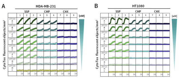
Figure 2. Discrimination of cytotoxic and cytostatic compounds. 96-well microplate graph showing the kinetic measurement of cell death as determined
by CytoTox green staining in response to several concentrations of SSP, CMP, and CHX in MDA-MB-231 cells (A) and HT-1080 cells (B).
[참고] 위 이미지에서 각각의 칸은 96-well 의 각 well 을 의미하며 각 칸 안의 자그만 그래프는 세포안 stained DNA 와 결합한 형광물질이 발생시키고 있는 형광의 세기를 나타내고 있습니다.
그래프가 수평이면 발생되는 형광이 없으므로 세포독성 반응이 없는 것을 의미하고 S자 그래프는 형광의 발생하고 있는 것을 의미하므로 세포독성 반응이 강하게 나타나고 있다는 것을 의미합니다.
96-well plate 에서 SSP 와 CMP 모두 concentration- dependent cytotoxic response 를 보였으며 세포에 따라 세포 독성정도는 약간 다르게 나타났다.
반면 CHX 는 두 개의 세포에 대해 모두 세포독성 반응이 나타나지 않았다 (Figure 2).

Figure 4. End point normalization of cytotoxic and cytostatic compounds. At the 72-hour end point, Triton
X-100 at a final concentration of 0.0625% was added to allow nuclear dsDNA staining by CytoTox green of all cells
present/well. The cytotoxic index was calculated by dividing the number of CytoTox green fluorescent objects by the total number of
DNA containing objects (fluorescent objects counted post Triton X-100 treatment).
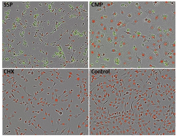
Figure 5. Morphological images. Phase-contrast and fluorescent images of NucLight HT-1080 showing morphological changes in response to SSP, CMP and CHX
Figure 4 는 실험 마지막 단계에서 MDA-MB-231, HT-1080 두 개의 세포에서 SSP, CMP, 그리고 CHX 3개의 물질의 세포독성 유도 정도를 수치화하여 나타낸 그래프이고
Figure 5 는 SSP, CMP, CHX 를 처리한 HT-1080 세포의 형광 이미지이다.
위 데이터로 부터 우리는 CytoTox Reagents 를 이용하는 IncuCyte® Live-Cell Analysis System 을 이용하면 Cytotoxicity 를 정확하게, 편리하게, 실시간으로, 시각적으로, 정량적으로 관찰할 수 있다는 것을 확인할 수 있다.
|
|
|
|